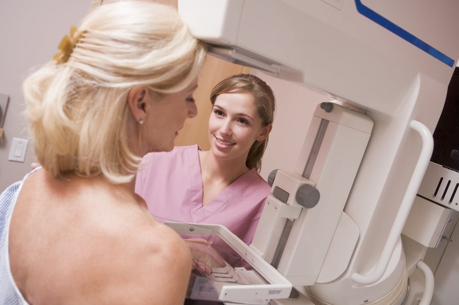
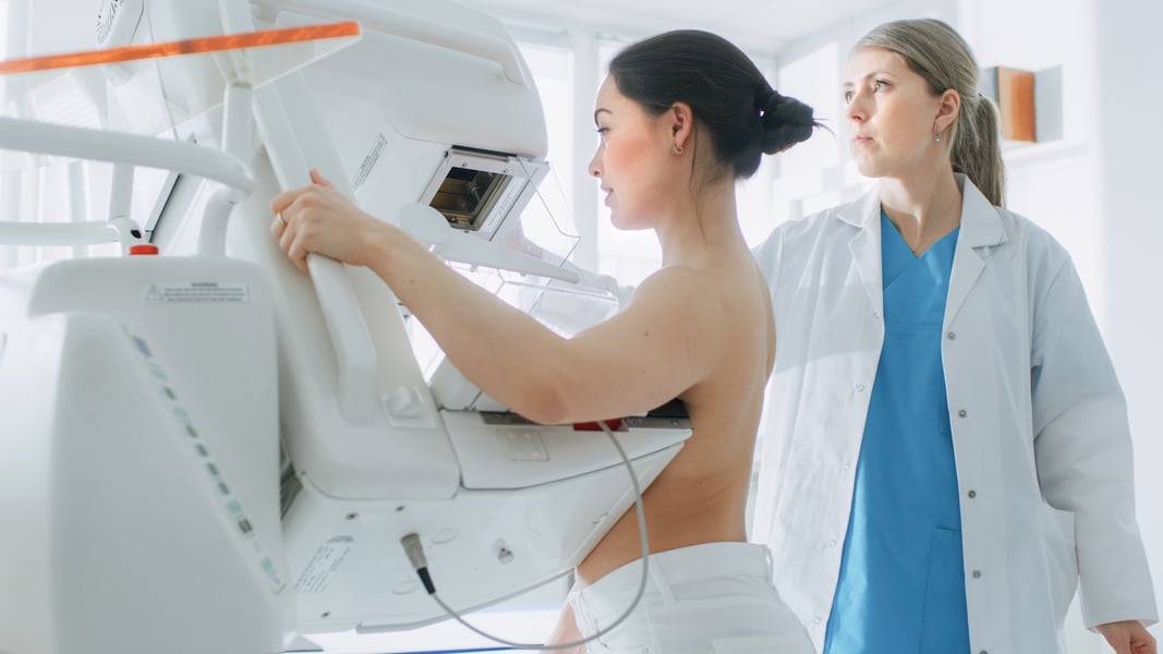
It's important that you have regular breast exams by your doctor since there are very few noticeable symptoms of breast cancer. Most women should start a screening mammogram around the age of 40. The exact time your doctor suggests you start will depend on your family history.
Breast cancer treatment is easier and more successful when breast cancer is detected early which is why it's so important to get your annual mammogram!
Watch the video below for a guide to breast cancer screening.
Clinical Breast Exams
During a clinical breast exam, your health care provider checks your breasts. You may be asked to raise your arms over your head, let them hang by your sides, or press your hands against your hips.
Your health care provider looks for differences in size or shape between your breasts. The skin of your breasts is checked for a rash, dimpling, or other abnormal signs. Your nipples may be squeezed to check for fluid.
Using the pads of the fingers to feel for lumps, your health care provider checks your entire breast, underarm, and collarbone area. A lump is generally the size of a pea before anyone can feel it. The exam is done on one side and then the other. Your health care provider checks the lymph nodes near the breast to see if they are enlarged.
If you have a lump, your health care provider will feel its size, shape, and texture. Your health care provider will also check to see if the lump moves easily. Benign lumps often feel different from cancerous ones. Lumps that are soft, smooth, round, and movable are likely to be benign. A hard, oddly shaped lump that feels firmly attached within the breast is more likely to be cancer, but further tests are needed to diagnose the problem.

What Every Woman Should Know About Breast Imaging
Other Imaging Tests
If an abnormal area is found during a clinical breast exam or with a mammogram, the doctor may order other imaging tests:
- Ultrasound: A woman with a lump or other breast change may have an ultrasound test. An ultrasound device sends out sound waves that people can’t hear. The sound waves bounce off breast tissues. A computer uses the echoes to create a picture. The picture may show whether a lump is solid, filled with fluid (a cyst), or a mixture of both. Cysts usually are not cancer. But a solid lump may be cancer.
- MRI: MRI uses a powerful magnet linked to a computer. It makes detailed pictures of breast tissue. These pictures can show the difference between normal and diseased tissue.
Breast Biopsy if Cancer is Supsected
A biopsy is the removal of tissue to look for cancer cells. A biopsy is the only way to tell for sure if cancer is present.
You may need to have a breast biopsy if an abnormal area is found during an exam or on a mammogram. An abnormal area may be felt during a clinical breast exam, but not seen on a mammogram. Or an abnormal area could be seen on a mammogram, but not be felt during a clinical breast exam. In this case, doctors can use imaging procedures (such as a mammogram, an ultrasound, or MRI) to help see the area and remove tissue.
Your doctor may refer you to a surgeon or breast disease specialist for a biopsy. The surgeon or doctor will remove fluid or tissue from your breast in one of several ways:
- Fine-needle aspiration biopsy: Your doctor uses a thin needle to remove cells or fluid from a breast lump.
- Core biopsy: Your doctor uses a wide needle to remove a sample of breast tissue.
- Skin biopsy: If there are skin changes on your breast, your doctor may take a small sample of skin.
- Surgical biopsy: Your surgeon removes a sample of tissue.
- An incisional biopsy takes a part of the lump or abnormal area.
- An excisional biopsy takes the entire lump or abnormal area.
A pathologist will check the tissue or fluid removed from your breast for cancer cells to create a pathology report. This report determines if cancer cells are found and gives the pathologist information about what type of breast cancer it is.
The video below discusses the facts about a breast biopsy. Watch it to learn more about what a breast biopsy is, the different types of breast biopsies, the reasons for needing one, and what to expect after the biopsy.

Lab Tests with Breast Tissue
If you are diagnosed with breast cancer, your doctor may order special lab tests on the breast tissue that was removed:
- Hormone receptor tests: Some breast tumors need hormones to grow. These tumors have receptors for the hormones estrogen, progesterone, or both. If the hormone receptor tests show that the breast tumor has these receptors, then hormone therapy is most often recommended as a treatment option. See the Hormone Therapy section.
- HER2/neu test: HER2/neu protein is found on some types of cancer cells. This test shows whether the tissue either has too much HER2/neu protein or too many copies of its gene. If the breast tumor has too much HER2/neu, then targeted therapy may be a treatment option. See the Targeted Therapy section.
It may take several weeks to get the results of these tests. The test results help your doctor decide which cancer treatments may be options for you.