Skin cancer is the most common form of cancer in the United States, with more people being diagnosed each year than all other cancers combined. Knowing what to look for can help catch it early when it’s much easier to treat.
The Importance of Skin Cancer Self-Examination
When detected early, skin cancer is almost always curable. This is why getting to know your skin through regular self-exams is so important so that any new or changing marks or lesions can be caught quickly.
Lesions, ulcers, or tumors on the skin should be checked out by a skin cancer specialist right away.
Marks and moles should be documented and monitored for changes during your self-exams. The Skin Cancer Foundation recommends head-to-toe self-examinations of the skin once a month and an annual exam by a dermatologist once a year.
How to Check Your Skin
Checking your skin means taking note of all the spots on your body. Spots typically include freckles, moles, birthmarks, age spots, bumps, sores, scabs, open wounds that bleed, and scaly patches.
For your self-exam, you’ll need a full-length mirror, a hand mirror, bright lighting, and a place to record your findings. When possible, ask someone to help check hard-to-see places.
- Examine your body front and back in the mirror, then look at the right and left sides with your arms raised. Women should lift their breasts to view the undersides.
- Bend elbows and look carefully at forearms, underarms, and palms. Also, check between fingers and under fingernails.
- Look at the backs of your legs and feet, between your toes, and the soles of your feet.
- Check the back of your neck and scalp with a hand mirror. Part the hair to get a closer look. A hair dryer may be helpful in raising your hair so it’s easier to see.
- Examine your back and buttocks with a hand mirror.
Sun exposure isn’t the only risk factor when it comes to developing skin cancer; that is why it’s important to examine all of your skin, including places that aren’t often (or ever) exposed to the sun or UV rays.
Resources for Keeping Track of Skin Changes
After the exam, it’s important to take note of your findings. An easy and effective way to do this is by downloading a Body Map to track new spots or changes in existing spots. On a printed diagram of the body, you simply make marks that correspond to the marks on your skin, then draw lines out to the margin to record the approximate size, color, and date. Use the same map to record your findings and compare them each month.
Smartphone apps like SkinVision and MySkinPal can also be used to track and take pictures of spots. It is important to remember, though, that these apps should never be used for diagnosis.
With each self-exam, you’ll become more familiar with what is normal for you, so anything unusual will draw your attention quickly, and you can have it checked out by your primary care physician or dermatologist if you have one.
What to Look For on Your Skin
It can be challenging to identify which marks on your skin are normal and which are not, especially if you’re prone to freckles or moles. The American Cancer Society recommends that you be on the lookout for an “ugly duckling” on your skin — any mark that looks different than all the others.
Additionally, the following ABCDE rule is helpful in spotting potential melanomas.
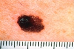
A for Asymmetry: Half of the mole or mark doesn’t match the other half.

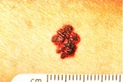
B for Border: Irregular, jagged, blurry, or notched edges.

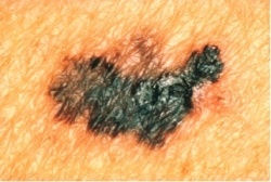
C for Color: Non-uniform color that includes different shades of black, brown, red, white, pink or blue patches.

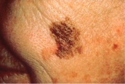
D for Diameter: The growth is more than ¼ inch in diameter (about the size of a pencil eraser.)

E for Evolving: The mole is growing or changing color or shape.
Not all skin cancers follow these rules, but many do. When in doubt about any mark on your skin that seems unusual, be cautious and have it looked at by a dermatologist.
Related reading: What is the Difference in Precancerous Skin Growths and Skin Cancer?
Detecting Skin Cancer
Typically, skin cancer detection involves a general practitioner or a dermatologist identifying an unusual area on the skin. However, they cannot definitively determine if the abnormal area is cancer until it is removed and tested, a procedure known as a biopsy. A biopsy is the most reliable method for diagnosing skin cancer.
In certain instances, a healthcare provider may opt to remove a seemingly harmless mole instead of allowing it to develop to determine if it is cancerous.
The skin biopsy procedure can take place in a medical office or as an outpatient procedure in a clinic or hospital. The choice of location depends on the size and location of the abnormal skin area, and local anesthesia is typically administered.
Once a skin cancer diagnosis is confirmed, the next step involves assessing its extent, a process known as staging. Staging skin cancer helps determine the appropriate treatment plan tailored to the patient's specific requirements. If the cancer has penetrated deeply into the skin or spread to lymph nodes, additional treatments are necessary to make sure all of the cancer cells have been removed following surgery. Depending on the stage, your oncologist will determine the necessary skin cancer treatments and create a personalized plan.
The skin cancer doctors at Compass Oncology offer the latest treatment options for melanoma and non-melanoma skin cancer in Portland-Vancouver.
Photography Sources:
- Figure 1: Unknown Photographer. (1988). Asymmetrical Melanoma. [Digital Image]. Skin Cancer Foundation.
- Figure 2: Unknown Photographer. (1988). Melanoma with Uneven Border. [Digital Image]. Skin Cancer Foundation.
- Figure 3: Unknown Photographer. (1988). Melanoma with Color Differences. [Digital Image]. Skin Cancer Foundation.
- Figure 4: Unknown Photographer. (1988). Melanoma with Diameter Change. [Digital Image]. Skin Cancer Foundation.


