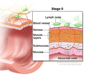
Stage 0 Colorectal Cancer
Also called carcinoma in situ, cancer is found only in the innermost lining of the colon or rectum.
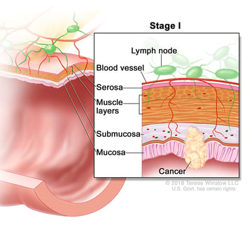
Stage I Colorectal Cancer
The tumor has grown into the inner wall of the colon or rectum. The tumor has not grown through the wall (T1 or T2, N0, M0).
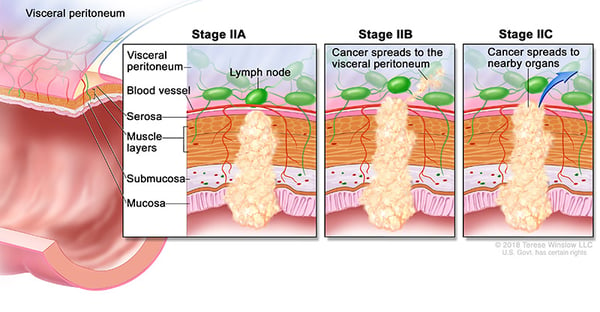
Stage II Colorectal Cancer
Stage IIA: The cancer has grown through the wall of the colon or rectum but has not spread to nearby tissue or to the nearby lymph nodes (T3, N0, M0).
Stage IIB: The cancer has grown through the layers of the muscle to the lining of the abdomen, called the visceral peritoneum. It has not spread to the nearby lymph nodes or elsewhere (T4a, N0, M0).
Stage IIC: The tumor has spread through the wall of the colon or rectum and has grown into nearby structures. It has not spread to the nearby lymph nodes or elsewhere (T4b, N0, M0).
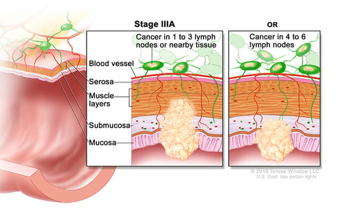
Stage III Colorectal Cancer
Stage IIIA: The cancer has grown through the inner lining or into the muscle layers of the intestine. It has spread to 1 to 3 lymph nodes or to a nodule of tumor cells in tissues around the colon or rectum that do not appear to be lymph nodes but has not spread to other parts of the body (T1 or T2, N1 or N1c, M0; or T1, N2a, M0).
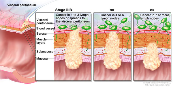
Stage IIIB: The cancer has grown through the bowel wall or to surrounding organs and into 1 to 3 lymph nodes or to a nodule of tumor in tissues around the colon or rectum that do not appear to be lymph nodes. It has not spread to other parts of the body (T3 or T4a, N1 or N1c, M0; T2 or T3, N2a, M0; or T1 or T2, N2b, M0).
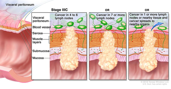
Stage IIIC: The cancer of the colon, regardless of how deep it has grown, has spread to 4 or more lymph nodes but not to other distant parts of the body (T4a, N2a, M0; T3 or T4a, N2b, M0; or T4b, N1 or N2, M0).
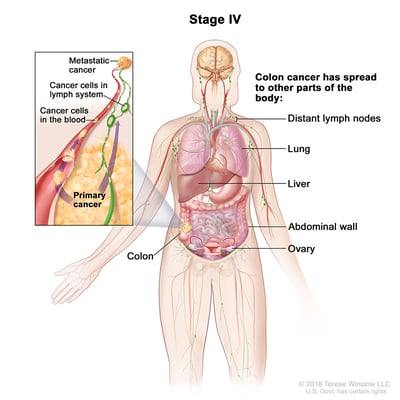
Stage IV Colorectal Cancer
Stage IVA: The cancer has spread to a single distant part of the body, such as the liver or lungs (any T, any N, M1a).
Stage IVB: The cancer has spread to more than 1 part of the body (any T, any N, M1b).
Stage IVC: The cancer has spread to the peritoneum. It may also have spread to other sites or organs (any T, any N, M1c).
Colorectal Cancer Care in the Portland-Vancouver Area
If you have been newly diagnosed with colorectal cancer, we encourage you to schedule an appointment with an oncologist. We offer personalized colorectal cancer treatment plans and second opinions for patients who live in the Portland, OR, or Vancouver, WA area. Find a colon and rectal cancer specialist near you.
